Your Elodea plant microscope images are ready in this website. Elodea plant microscope are a topic that is being searched for and liked by netizens today. You can Get the Elodea plant microscope files here. Find and Download all royalty-free vectors.
If you’re looking for elodea plant microscope images information connected with to the elodea plant microscope topic, you have come to the right blog. Our website frequently gives you hints for seeking the highest quality video and picture content, please kindly hunt and find more informative video articles and images that fit your interests.
Elodea Plant Microscope. A small leaf has been removed from the plant and placed with the lower surface down in a drop of water on a microscope slide. Plastids are organelles characteristic of plant cells they are clearly differentiated protoplasmic bits of special plant cell structure and function organelles. Create a wet mount using the elodea leaf tip. These leaves are two cells thick, so you should be able to focus up and down to see that the cells in one layer are larger than those in the other.
 elodea cytoplasmic streaming YouTube From youtube.com
Observe the cells under normal conditions, and make a sketch of what you see. Pond water was mixed with the leaf sample so there are some organisms interacting with the lea. Ad mvx10 macroview microscope for efficient, bright fluorescence imaging. Only one layer of cells is in focus when using the high. It can grow in aquariums, and it is an easy specimen to study under a microscope as an example of a plant cell. Ad mvx10 macroview microscope for efficient, bright fluorescence imaging.
Elodea leaf cell under microscope plant cell cells worksheet lab activities.
When studying an elodea cell under a microscope, it is important to remember that the cell consists of two layers, yet only one of them can be in focus. To find specimens using low, medium, and high power. Plant cells vs animal cells with diagrams animal cell plant cell diagram plant cell. Examining elodea (pondweed) under a compound microscope. Carefully cut the “growing end’ from the tip of an elodea leaf. Leaf cells elodea is a decorative aquatic plant often found in fish tanks.
 Source: youtube.com
Elodea leaf cell under microscope plant cell cells worksheet lab activities. Name:_____ elodea plant leaf & onion skin microscope lab period: To observe onion (bulb portion of the plant) and elodea (a common aquarium plant) cells? As you can see in the image, the shapes of the cells vary to some degree, so taking an average of three cells’ dimensions, or even the results from the entire class, gives a more accurate determination of “typical” elodea cell size. To make a wet mount slide.
 Source: youtube.com
A small leaf has been removed from the plant and placed with the lower surface down in a drop of water on a microscope slide. To view your own (or your partner’s). Leaf cells elodea is a decorative aquatic plant often found in fish tanks. To prepare a sample for observation,. Examining elodea (pondweed) under a compound microscope.
 Source: app.emaze.com
These pigments trap the energy from solar light, which is used by the plants for the process of photosynthesis to. Lab manual exercise 1 lab manual exercise 1 elodea plant cells at 40x 100x 400x lab manual exercise 1. Leaf cells elodea is a decorative aquatic plant often found in fish tanks. Only one layer of cells is in focus when using the high. Microscopic video of an elodea leaf at three separate powers.
 Source: fineartamerica.com
Ad mvx10 macroview microscope for efficient, bright fluorescence imaging. Wolfe® advanced led series binocular microscope with 4 objectives item #591004 $795.00 quick view wolfe® cfl educational microscope item #590950 $279.00 Using one small leaf of elodea, prepare a wet mount elodea leaf plant cell under the microscope plant cell things cm as the slide warms from the light of the microscope, you may see the chloroplasts moving, a process called cytoplasmic streaming these are often very. Get a single leaf from the elodea plant and mount it on a slide, cover it with a drop of water and a cover slip. Observe the cells under normal conditions, and make a sketch of what you see.
 Source: schematron.org
Get a single leaf from the elodea plant and mount it on a slide, cover it with a drop of water and a cover slip. The elodea leaf is composed of two layers of cells. The elodea plant is traded under many names including anacharis elodea densa and brazilian waterweed. High schoolers observe the general structure and organelles of plant and animal cells. Microscopic video of an elodea leaf at three separate powers.
 Source: mybiology230.blogspot.com
(this piece should be about 5 mm in size.) place the elodea leaf tip onto a drop of water that has been placed on a blank slide. Reproduces primarily through stem fragments. Beginners and experts alike should feel free to post anything that helps people learn more about plants and. As a result, only part of constituent parts of the cell will be visible. Elodea leaf under microscope 40x labeled.
 Source: youtube.com
Beginners and experts alike should feel free to post anything that helps people learn more about plants and. The elodea leaf is composed of two layers of cells. Ad mvx10 macroview microscope for efficient, bright fluorescence imaging. 40x 400x compound monocularbiological microscope45 degree angled headelectric lightedbeginner slides plant cell things under a microscope plant cell picture. Note that the mpeg4 video format reduces the video time from 3 minutes to 1 minute and creates a time lapse effect.
 Source: microspedia.blogspot.com
Elodea leaf cell under microscope plant cell cells worksheet lab activities. When studying an elodea cell under a microscope, it is important to remember that the cell consists of two layers, yet only one of them can be in focus. Beginners and experts alike should feel free to post anything that helps people learn more about plants and. To observe onion (bulb portion of the plant) and elodea (a common aquarium plant) cells? It can grow in aquariums, and it is an easy specimen to study under a microscope as an example of a plant cell.
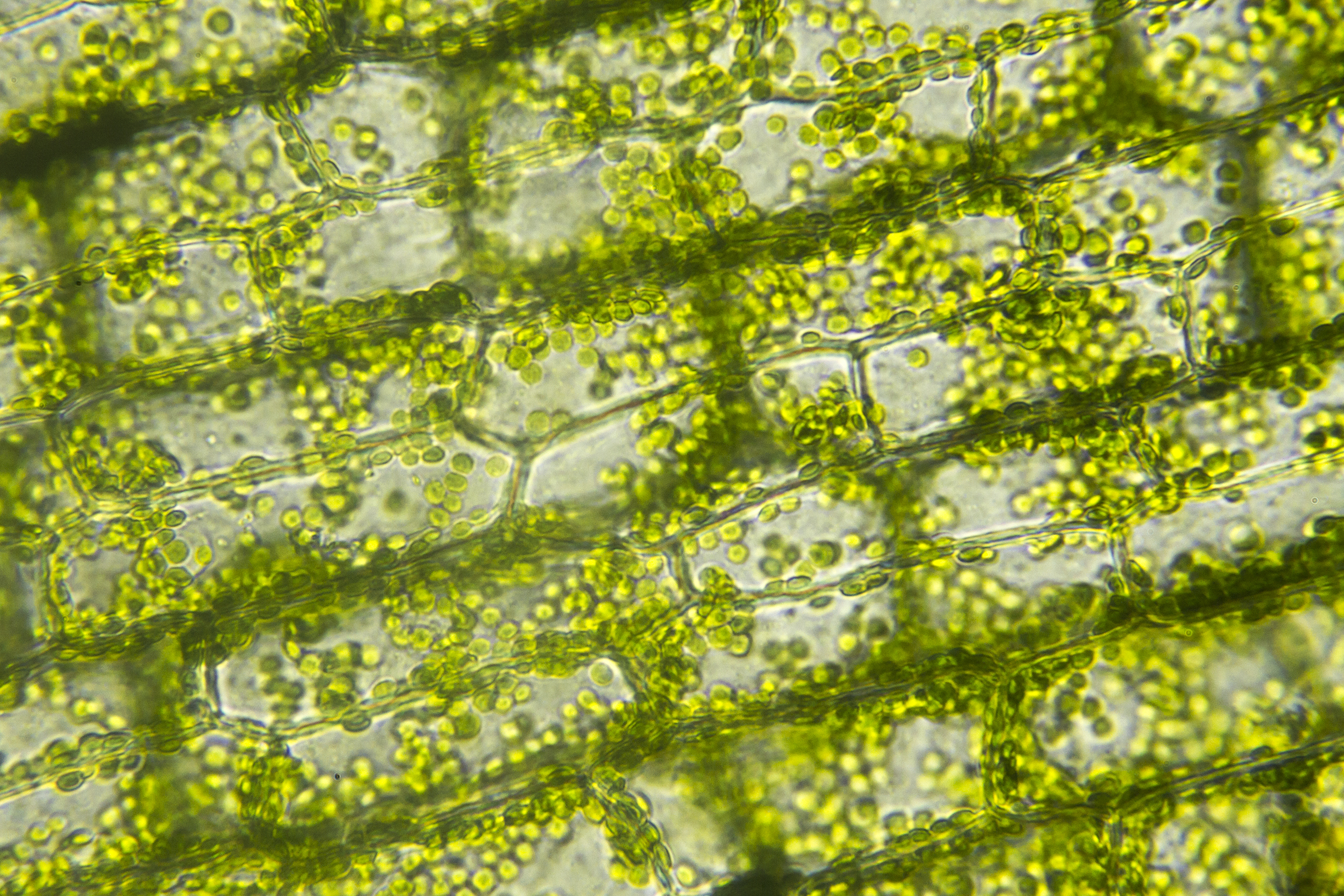
To observe onion (bulb portion of the plant) and elodea (a common aquarium plant) cells? To prepare a sample for observation,. It can grow in aquariums, and it is an easy specimen to study under a microscope as an example of a plant cell. Ad mvx10 macroview microscope for efficient, bright fluorescence imaging. Elodea leaf cell under microscope plant cell cells worksheet lab activities.

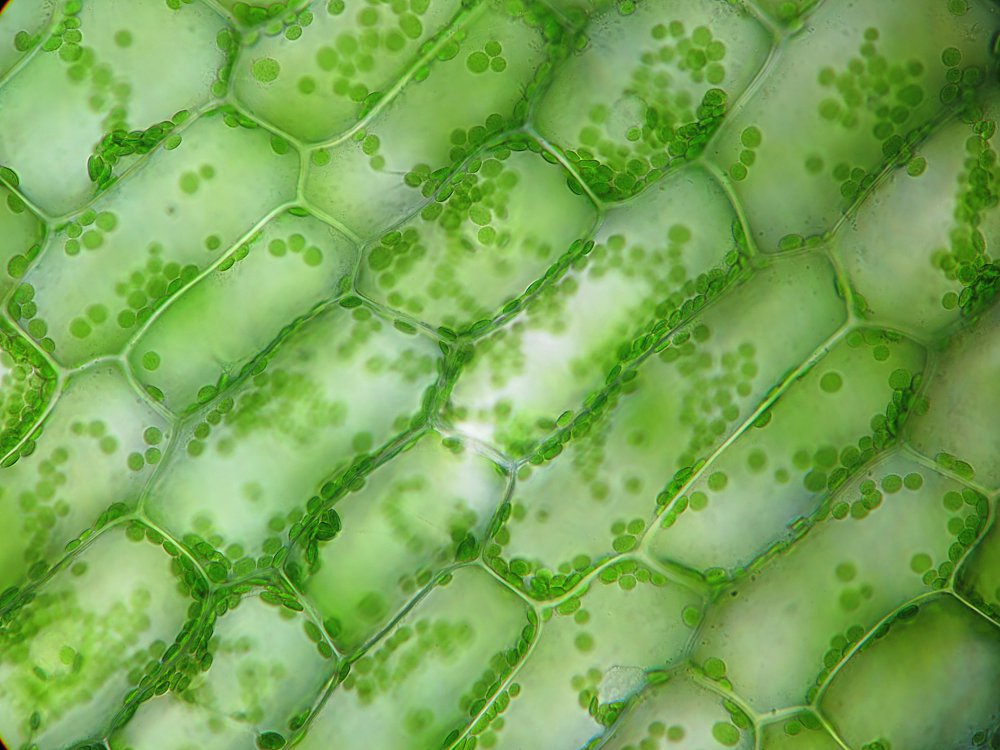
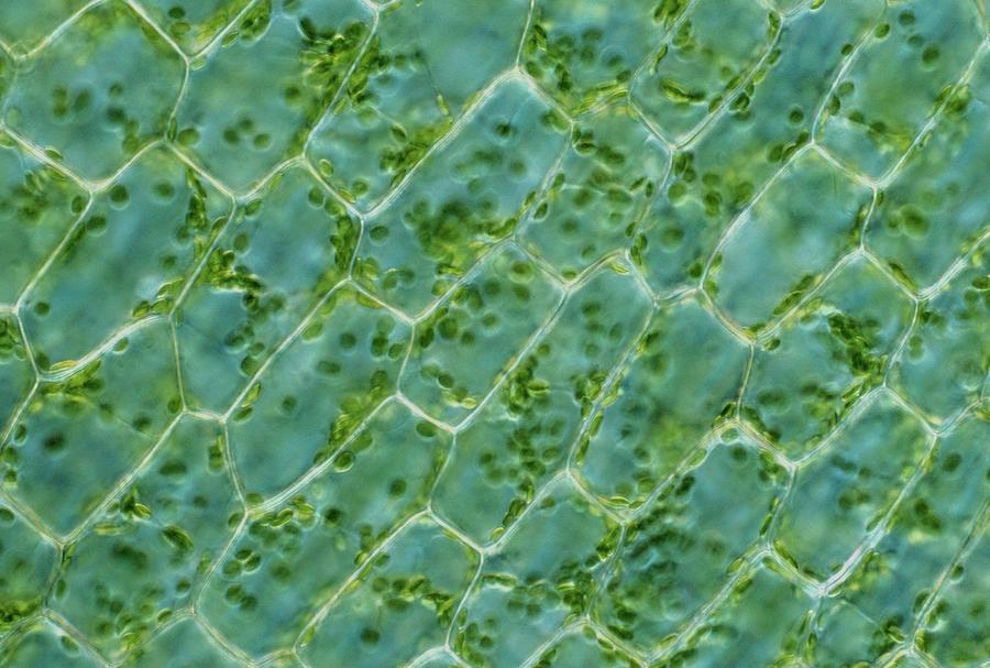
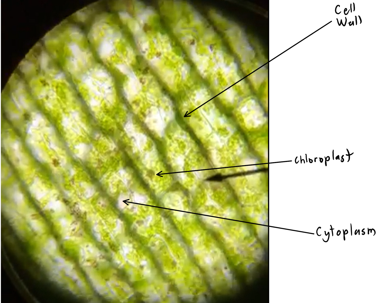
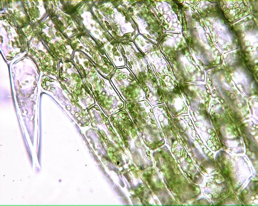
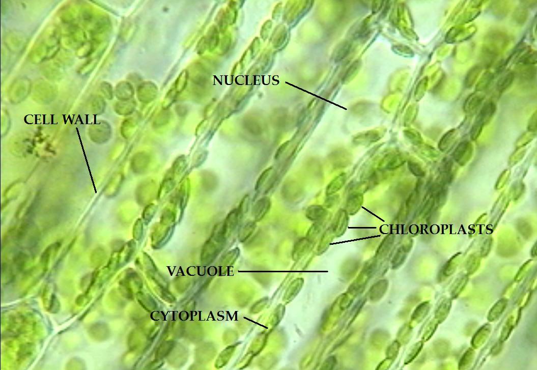
Ad mvx10 macroview microscope for efficient, bright fluorescence imaging. The green disks in elodea plant cell under a microscope represent specialized plant cell organelles, known as chloroplasts. Plastids are organelles characteristic of plant cells they are clearly differentiated protoplasmic bits of special plant cell structure and function organelles. Write two sentences about what you already know about plant cells. In this lesson, students microscopically observe various subcellular components and.
 Source: alamy.com
See information on suppliers here. To prepare a sample for observation,. Elodea leaf cell under microscope plant cell cells worksheet lab activities. A cells plant cell parts cell parts photosynthesis and cellular respiration. Pond water was mixed with the leaf sample so there are some organisms interacting with the lea.
 Source: schematron.org
Table diagram your microscope observations in the circles. In this lesson, students microscopically observe various subcellular components and. This sub is a place for the discussion of botany at any level. A “typical” elodea cell is approximately 0.05 millimeters long (50 micrometers long) and 0.025 millimeters wide (25 micrometers wide). When studying an elodea cell under a microscope, it is important to remember that the cell consists of two layers, yet only one of them can be in focus.
 Source: youtube.com
Chloroplasts moving by cytoplasmic streaming in the cells of aquatic plant elodea you. To find specimens using low, medium, and high power. The elodea leaf is composed of two layers of cells. To view your own (or your partner’s). Carefully cut the “growing end’ from the tip of an elodea leaf.
 Source: youtube.com
Carefully cut the “growing end’ from the tip of an elodea leaf. Beginners and experts alike should feel free to post anything that helps people learn more about plants and. To view your own (or your partner’s). To make a wet mount slide. Lab 2 microscopes and cells gen bio 1 flashcards questions answers quizlet.
 Source: youtube.com
Elodea leaf cell under microscope plant cell cells worksheet lab activities. Pick off an entire healthy looking elodea leaf, with fingers or small scissors and place it on the microscope slide. Table diagram your microscope observations in the circles. Chloroplasts moving by cytoplasmic streaming in the cells of aquatic plant elodea you. Wolfe® advanced led series binocular microscope with 4 objectives item #591004 $795.00 quick view wolfe® cfl educational microscope item #590950 $279.00
 Source: youtube.com
As you can see in the image, the shapes of the cells vary to some degree, so taking an average of three cells’ dimensions, or even the results from the entire class, gives a more accurate determination of “typical” elodea cell size. A cells plant cell parts cell parts photosynthesis and cellular respiration. Examining elodea (pondweed) under a compound microscope. Elodea leaf under microscope 40x labeled. Write two sentences about what you already know about plant cells.
 Source: microspedia.blogspot.com
Cover the specimen with a cover slip. Only one layer of cells is in focus when using the high. Pond water was mixed with the leaf sample so there are some organisms interacting with the lea. As a result, only part of constituent parts of the cell will be visible. Reproduces primarily through stem fragments.
 Source: youtube.com
Examining elodea (pondweed) under a compound microscope. Pick off an entire healthy looking elodea leaf, with fingers or small scissors and place it on the microscope slide. Ad mvx10 macroview microscope for efficient, bright fluorescence imaging. The elodea leaf is composed of two layers of cells. Place the slide onto the microscope state and observe at the leaf under the microscope.
This site is an open community for users to share their favorite wallpapers on the internet, all images or pictures in this website are for personal wallpaper use only, it is stricly prohibited to use this wallpaper for commercial purposes, if you are the author and find this image is shared without your permission, please kindly raise a DMCA report to Us.
If you find this site beneficial, please support us by sharing this posts to your preference social media accounts like Facebook, Instagram and so on or you can also save this blog page with the title elodea plant microscope by using Ctrl + D for devices a laptop with a Windows operating system or Command + D for laptops with an Apple operating system. If you use a smartphone, you can also use the drawer menu of the browser you are using. Whether it’s a Windows, Mac, iOS or Android operating system, you will still be able to bookmark this website.


